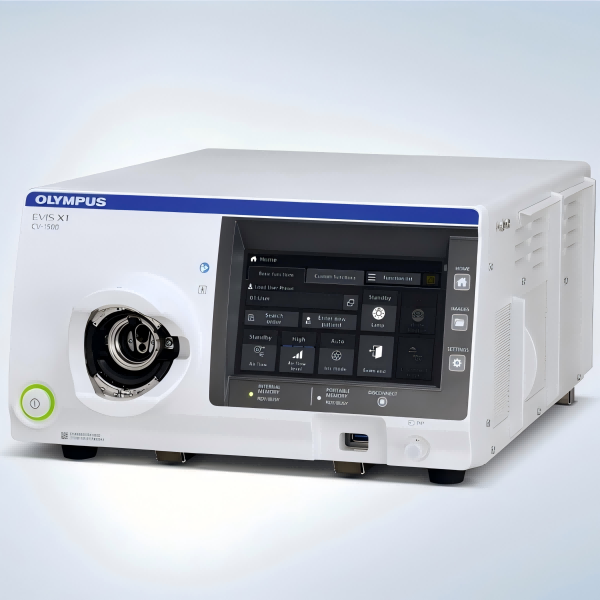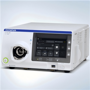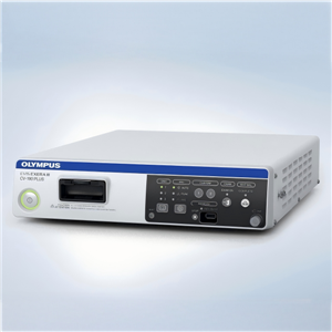
Olympus CV-190 PLUS Video Processor


- Olympus
- Japan
Olympus EVIS EXERA III CV-190 PLUS Video Processor
Video Processing Powering Advanced Endoscopy
Overview
Olympus EVIS EXERA III CV-190 PLUS Video Processor
Video Processing Powering Advanced Endoscopy
Main Features
· NBI (Narrow Band Imaging) in EVIS EXERA III 190 Series scopes provides twice the viewable distance of EVIS
· EXERA II 180 Series scopes and offers much greater contrast between blood vessels and mucosa.
· CV-190 PLUS contains the electronics to operate the dual focus that delivers an optimal view whether close up or distant by connecting HQ scopes.
· The newly designed waterproof one-touch connector enables a one-step connection to the light source and does not require a separate scope cable for the video processor.
· A new and improved image processing delivers sophisticated image quality via enhanced color reproduction,minimized image noise, and reduced halation.
· The pre-freeze function selects the clearest still image automatically, saving time.
· Compatible with GI/BF endoscopes of EVIS 100/130/140/150 Series, EVIS EXERA 160/145 Series,EVIS EVERA II 180/165 Series, EVIS EXERA III 190/185 Series, and OPTERA 170 Series.
· 16:9 and 16:10 output for a HDTV monitor is available.Compatible with analog, HD-SDI, and DVI output.
· Link connection to peripheral devices avoids complicated cable connections and accelerates transmission speed.
· OLYMPUS documentation system enhances networking expandability.
· Picture-in-picture and index function effectively enhance your observation.
· Portable memory is compatible, which is standard for data management. Simply connect and upload.
· Supports DV output to compatible documentation devices.
Specifications
Power Supply | Voltage | 100-240 V AC (NTSC)/220-240 V AC (PAL); within ±10% |
Frequency 5 | 0/60 Hz; within ±1 Hz | |
Consumption electric power | 150 VA | |
Size | Dimensions (W x H x D) | 370 x 85 x 455 mm; 382 x 91 x 489 mm (maximum) |
Weight | 10.7 kg | |
Classification(medical electrical equipment) | Type of protection against electric shock | Class I |
Degree of protection against electric shock of applied part | Depend on applied part. See also applied part (videoscope). | |
Degree or protection against explosion | The video system center should be kept away from flammable gases. | |
Observation | Analog HDTV signal output | Either RGB (1080/60I: NTSC)/(1080/50I: PAL) or YPbPr (1080/60I: NTSC)/(1080/50I: PAL)output can be selected. |
Analog SDTV signal output | VBS composite (480/60I: NTSC)/(576/50I: PAL), Y/C (480/60I: NTSC)/(576/50I: PAL), andRGB(480/60I: NTSC)/(576/50I: PAL); simultaneous outputs possible. | |
Digital signal output | HD-SDI (SMTPE 292M), SD-SDI (SMPTE 259M), DV (IEEE 1394), and DVI (WUXGA, 1080p or SXGA) can be selected. | |
White balance adjustment | White balance adjustment is possible using the white balance button on the front panel. | |
Standard color chart output | The “Color bar” or the “50% white” screen can be displayed. | |
Color tone adjustment | The following color tone adjustments are possible using the color tone level adjustment button and color tone selector button on the keyboard. | |
Automatic gain control (AGC) | The image can be electronically amplified when the light is inadequate due to the distal end of the endoscope being too far from the object. | |
Contrast | N (Normal): Normal image | |
Iris | The auto iris modes can be selected using the “iris mode” switch on the front panel. | |
Image enhancement setting | Fine patterns or edges in the endoscopic images can be enhanced electrically to increase the image sharpness. | |
Switching the enhancement modes | The enhancement level can be selected from 3 levels (OFF, 1, 2, and 3) using the image enhancement mode button on the front panel. | |
Image size selection | The size of the endoscopic image can be changed using the “IMAGE SIZE” key on the keyboard. | |
Freeze | An endoscopic image is frozen using an endoscope or the “FREEZE” key on the keyboard. | |
Switching the method of freezing the endoscopic image | Pre-freezing: The image with the least blur is selected from the images captured in the set time period before freeze operation and displayed. | |
Fog free function | When a compatible endoscope is connected to the video system center, the fog free function can be used. | |
Endoscope’s remote switches function | The functions of the remote switches on the endoscope can be set in the user settings. | |
Reset to defaults | The following settings can be reset to their defaults using the reset button on the front panel. | |
Remote control | The following ancillary equipment can be controlled (specified models only).Monitor, DVR, Video printer, Image filing system | |
Documentation | Patient data | The following data can be displayed on the monitor using the keyboard. |
Displaying the record state | The recording state of the following ancillary equipment can be displayed on the monitor. | |
Displaying the image information | The following data can be displayed on the monitor. | |
Advance registration of patient data | Up to 50 patient’s data can be registered. Patient ID, Patient name, Sex and age, Date of birth | |
Portable Memory | Media | MAJ-1925 (OLYMPUS) |
Recording format | TIFF: no compression, JPEG (1/5): approx. 1/5 compression, JPEG (1/10): approx. 1/10 compression | |
Number of recording images | TIFF: approx. 227 images, JPEG (1/5): approx. 1024 images, JPEG (1/10): approx. 2048 images | |
Memory Backup | User settings | Up to 20 user settings can be registered. |
Memorization of selected setting | The following settings are held in memory even after the video system center is turned OFF. | |
Lithium battery | Life: 5 years |